While conventional CT imaging produces images with a slice thickness of roughly 1mm, MicroCT is capable of resolving detail in the micrometre (μm) range. This…
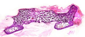
A Hematoxylin and eosin stained slide of a posterolateral fusion mass in an ovine model.
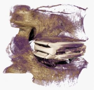
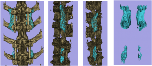
CT modeling is accomplished using a CT volume with a combination of 3DSlicer, Blender, and other 3D rendering software platforms. The image above is from…
