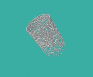
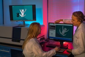
While conventional CT imaging produces images with a slice thickness of roughly 1mm, MicroCT is capable of resolving detail in the micrometre (μm) range. This…
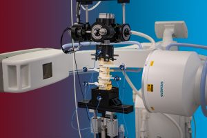
Fluoroscopy in the laboratory is an invaluable tool. Real-time fluoroscopic video allows us to gain a better understanding of the specific kinematic conditions occurring during…
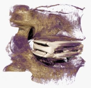
The image above was acquired from a 3D MicroCT volume, and represents a bioactive dowel used in an in-vivo investigational study of various biomaterials (ovine…
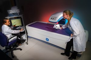
DEXA scanning allows the laboratory to assess bone quality prior to inclusion in cadaveric studies. This is especially important for studies requiring a degree of…
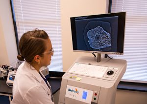
High resolution digital microradiography allows for detailed thin-section imaging.
The laboratories state-of-the-art micro CT system offers a new method for quantifying bone growth and conducting volumetric and surface area analysis of orthopaedic implant technology,…
