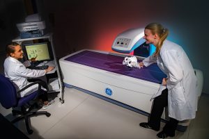
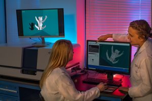
DEXA scanning allows the laboratory to assess bone quality prior to inclusion in cadaveric studies. This is especially important for studies requiring a degree of…
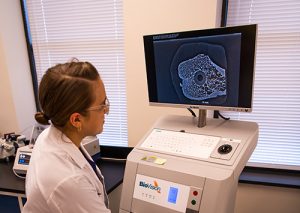
High resolution digital microradiography allows for detailed thin-section imaging.
The laboratories state-of-the-art micro CT system offers a new method for quantifying bone growth and conducting volumetric and surface area analysis of orthopaedic implant technology,…
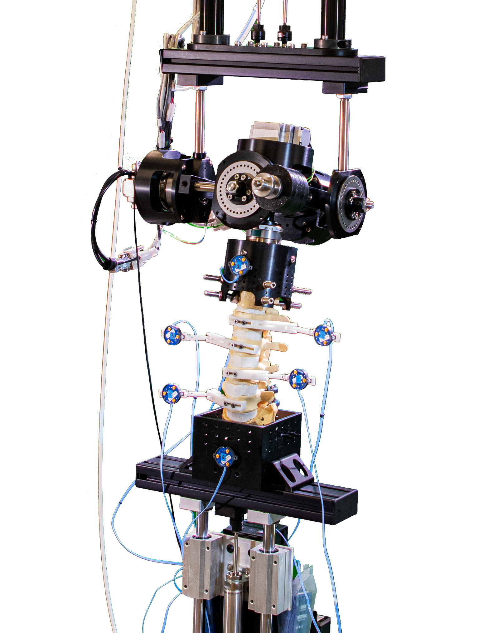
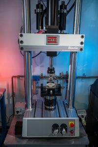
The laboratory is equipped with the latest MTS Bionix testing system in an axial/torsional configuration. Load data is captured through the systems load cell and…
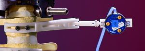
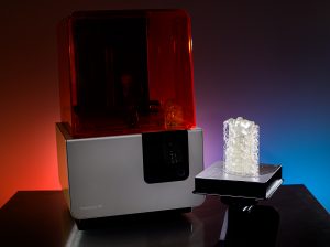
One example of how 3D printing and rapid prototyping has changed how the laboratory functions can be found with these 3D printed Radiolucent OptoTrak marker…
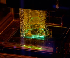
The laser of the labs Formlabs 3D printer is able to print distinct layers at a resolution of 50 microns. This insures the production of…
Rapid prototyping, preoperative models, and functional laboratory components, are all made possible with the labs 3D SLA printer.
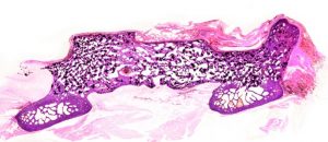
A Hematoxylin and eosin stained slide of a posterolateral fusion mass in an ovine model.
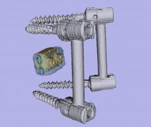
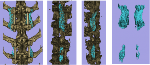
In-vivo interbody fusion cage location can be assessed in three dimensions with the help of CT modeling. Further investigation involving 3D printed models of the…
CT modeling is accomplished using a CT volume with a combination of 3DSlicer, Blender, and other 3D rendering software platforms. The image above is from…
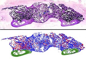
In this example, an H&E stained posterolateral fusion mass (PLF) is segmented into three areas: Bone, material, and transverse processes. Area calculations are subsequently obtained.
