Biomechanical Testing
6DOF Spine Simulator – MTS Bionix – Optotrak Motion Capture – Strain Measurements

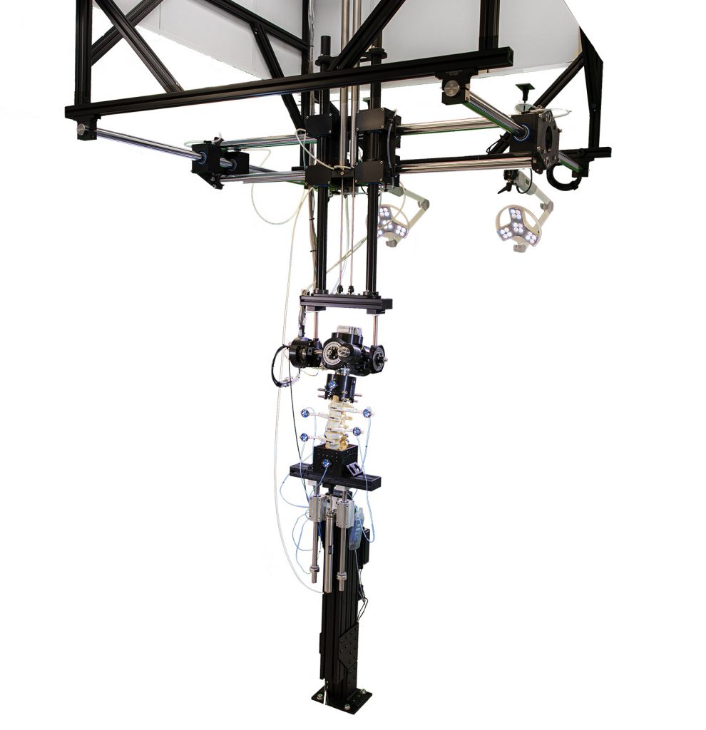
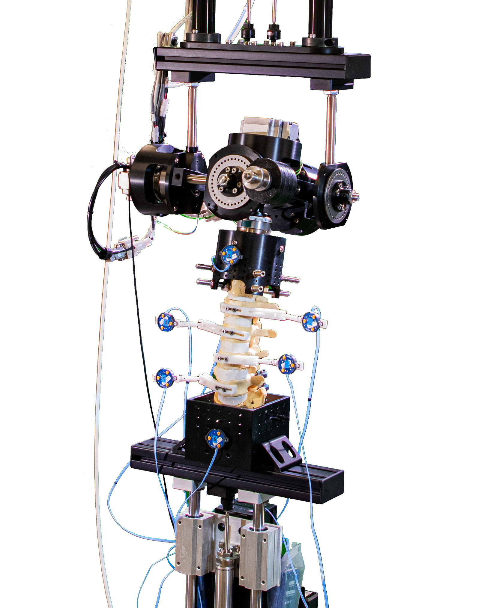
The centerpiece of the Orthopaedic Spinal Research Laboratory is the six degree of freedom (6DOF) spine simulator, designed by mechanical engineer, Jeff Gordon, MS. and Bryan Cunningham, PhD. The spine simulator recreates the natural movement of spinal kinematics, and is used to perform biomechanical assessments of spinal reconstruction techniques.
The six degree of freedom spine simulator can produce up to 64 Nm of flexion/extension, lateral bending, and/or axial rotation torques. Each of the three axes – X, Y and Z – is controlled independently to induce a torque profile, to lock, or to operate on bearings – allowing inherently coupled rotational spinal motions to occur unconstrained. Translations in all three axes – X, Y and Z – can be unconstrained or locked if required. This technology combined with advanced histological and radiographic laboratories permit basic scientific research in new areas such as dynamic spinal stabilization, which is intended to preserve spinal mobility versus traditional methods of spinal fusion.
The simulator also allows application of a follower load to simulate body weight and trunk loads. Segmental motion tracking is performed using an optoelectronic system (Optotrak, Northern Digital) which can record rotations to within 0.1 degree and translations to within 0.1 mm .
6 DOF Spine Simulator Video
The above video takes a specimen through a full range of motion test including flexion/extension, lateral bending, and axial rotation. Typically three cycles of each are recorded, however; this video uses only one cycle each for demonstration purposes.
Lateral Fluoroscopic View Of Flexion/Extension
A fluoroscopic image intensifier can provide real-time images during testing. The video below shows a lateral fluoroscopic view of flexion and extension being performed with the 6DOF spine simulator.
Real time fluoroscopic video provides a multitude of quantitative data, including: analysis of the integrity of each motion segment, unique spinal kinematic signature of each motion segment with respect to center of intervertebral rotation, and the effects of spinal destabilization / reconstruction on segmental range of motion and multi-directional flexibility.
Fluoroscopic video taken of specimens on the 6DOF Spine Simulator are used in combination with other data acquisition systems, including the Optotrak system and intradiscal pressure gauges, in order to quantify the kinematic multi-directional flexibility characteristics and segmental load distribution properties of the operative functional spinal units evaluated.
